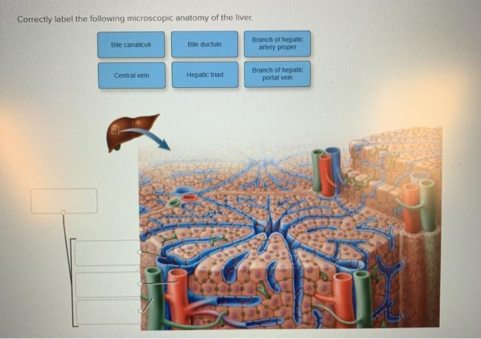
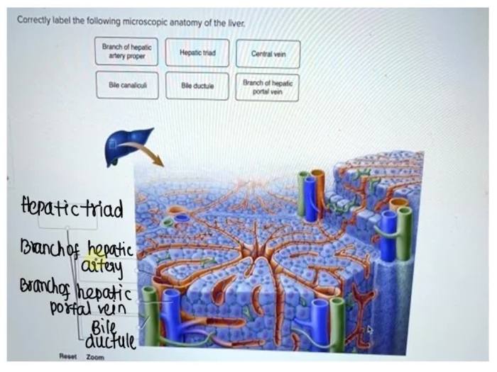
Correctly label the following microscopic anatomy of the liver. – Correctly labeling the microscopic anatomy of the liver is crucial for understanding its intricate structure and function. This guide provides a comprehensive overview of the cellular components, hepatic lobules, bile production, blood supply, and innervation of the liver, empowering readers with the knowledge to accurately identify and describe its microscopic features.
Liver Anatomy Overview: Correctly Label The Following Microscopic Anatomy Of The Liver.

The liver is the largest internal organ in the human body, weighing approximately 1.5 kilograms (3.3 pounds). It is located in the upper right quadrant of the abdominal cavity, beneath the diaphragm, and is responsible for a wide range of essential functions, including metabolism, detoxification, and bile production.The
liver is composed of two main lobes, the right and left lobes, which are further divided into eight segments. The liver parenchyma, the functional tissue of the liver, is made up of hexagonal lobules, which are the basic structural and functional units of the liver.
Microscopic Anatomy of the Liver

The liver is composed of several types of cells, including hepatocytes, Kupffer cells, and liver sinusoidal endothelial cells. Hepatocytes are the most abundant cells in the liver and are responsible for the majority of the liver’s functions, including metabolism, detoxification, and bile production.
Kupffer cells are macrophages that are located in the liver sinusoids and are responsible for removing bacteria and other foreign particles from the blood. Liver sinusoidal endothelial cells line the liver sinusoids and are responsible for the exchange of nutrients and waste products between the blood and the hepatocytes.
Hepatic Lobules
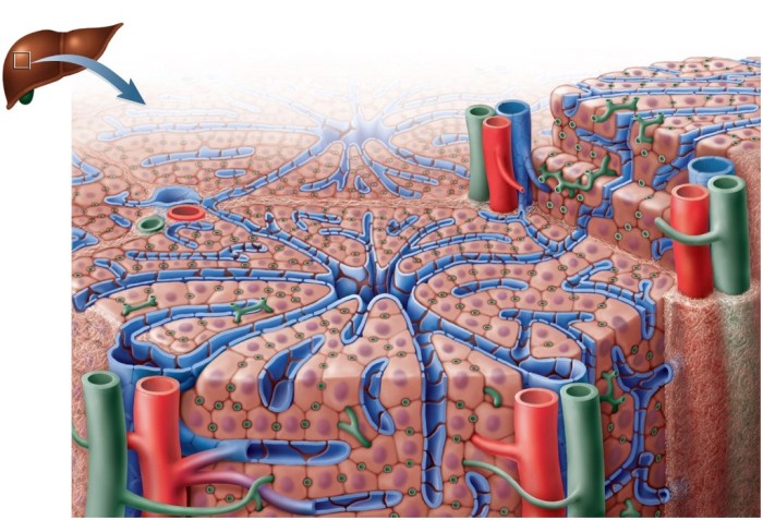
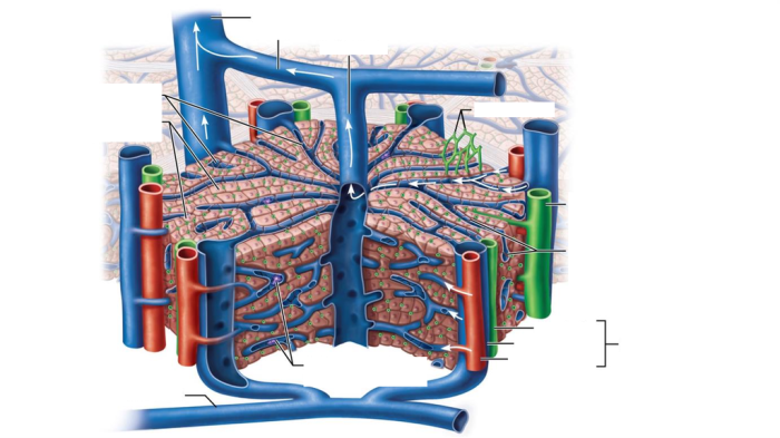
The liver is composed of hexagonal lobules, which are the basic structural and functional units of the liver. Each lobule is surrounded by a portal triad, which is composed of a hepatic artery, a portal vein, and a bile duct.
The hepatic artery supplies oxygenated blood to the liver, the portal vein supplies deoxygenated blood from the intestines, and the bile duct drains bile from the liver. The central vein runs through the center of each lobule and collects blood from the sinusoids.
Bile Production and Secretion
Bile is a fluid that is produced by the hepatocytes and is responsible for emulsifying fats and aiding in their digestion. Bile is produced in the hepatocytes and is then transported through the bile canaliculi, which are small channels that run between the hepatocytes.
The bile canaliculi merge to form bile ducts, which eventually drain into the common hepatic duct. The common hepatic duct joins with the cystic duct from the gallbladder to form the common bile duct, which drains bile into the duodenum.
Blood Supply to the Liver, Correctly label the following microscopic anatomy of the liver.
The liver receives a dual blood supply from the hepatic artery and the portal vein. The hepatic artery supplies oxygenated blood to the liver, while the portal vein supplies deoxygenated blood from the intestines. The hepatic artery and portal vein branch into smaller vessels within the liver, which eventually form the liver sinusoids.
The liver sinusoids are lined by liver sinusoidal endothelial cells, which are responsible for the exchange of nutrients and waste products between the blood and the hepatocytes.
Innervation of the Liver
The liver is innervated by the autonomic nervous system, which is responsible for regulating liver function. The sympathetic nervous system stimulates the liver to release glucose into the bloodstream, while the parasympathetic nervous system stimulates the liver to produce bile.
Expert Answers
What are the main cellular components of the liver?
Hepatocytes, Kupffer cells, and liver sinusoidal endothelial cells.
What is the functional unit of the liver?
The hepatic lobule.
How is bile produced and secreted?
Bile is produced by hepatocytes and secreted into bile canaliculi, which merge to form bile ducts.

